What is a visual evoked potential? (VEP)
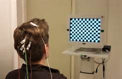
A visual evoked potential is an evoked potential caused by a visual stimulus, such as an alternating checkerboard pattern on a computer screen. Responses are recorded from electrodes that are placed on the back of your head and are observed as a reading on an electroencephalogram (EEG). These responses usually originate from the occipital cortex, the area of the brain involved in receiving and interpreting visual signals.
When is the VEP used?
A doctor may recommend that you go for a VEP test when you are experiencing changes in your vision that can be due to problems along the pathways of certain nerves. Some of these symptoms may include:
Loss of vision (this can be painful or non-painful);
Double vision;
Blurred vision;
Flashing lights;
Alterations in colour vision; or
Weakness of the eyes, arms or legs.
These changes are often too subtle or not easily detected on clinical examination in the doctor’s surgery. In general terms, the test is useful for detecting optic nerve problems. This nerve helps transfer signals to allow us to see, so testing the nerve allows the doctor to see how your visual system responds to light. The test is also useful because it can be used to check vision in children and adults who are unable to read eye charts.
What does the VEP detect?
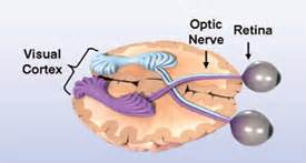
The VEP measures the time that it takes for a visual stimulus to travel from the eye to the occipital cortex. It can give the doctor an idea of whether the nerve pathways are abnormal in any way. For example, in multiple sclerosis, the insulating layer around nerve cells in the brain and spinal cord (known as the myelin sheath) can be affected. This means that it takes a longer time for electrical signals to be conducted from the eyes, resulting in an abnormal VEP. A normal VEP can be fairly sensitive in excluding a lesion of the optic nerve, along its pathways in the anterior part of the brain.
Visual evoked potential (VEP)The VEP is a standardised and reproducible test of optic nerve function
It is more sensitive compared to magnetic resonance imaging (MRI) in detecting lesions affecting the visual pathway in front of the optic chiasm (area in the optic pathway where the optic nerve crosses sides)
What the results of the VEP may show
The VEP is particularly useful in detecting past optic neuritis. This refers to inflammation of the optic nerve, associated with swelling and progressive destruction of the sheath covering the nerve, and sometimes the nerve cable. As the nerve sheath is damaged, the time it takes for electrical signals to be conducted to the eyes is prolonged, resulting in an abnormal VEP. This may be seen in multiple sclerosis – one of the most common causes of optic neuritis (as above). Abnormal VEP’s are seen in multiple sclerosis patients due to the presence of optic neuritis.
The following are less easily differentiated but may cause abnormal VEPs:
Optic neuropathy – this can be due to damage of the optic nerve from a number of causes, including: a blockage of the nerve’s blood supply, nutritional deficiencies, or toxins. As the nerve is damaged, electrical signals do not conduct properly. Examples include diabetes in the advanced stages which can be associated with damage to the blood vessels and nerves supplying the eyes, or toxic amblyopia which is a condition of the eyes associated with decreased vision, due to a toxic reaction in part of the optic nerve.
Tumours or lesions compressing the optic nerve – if the optic nerve is compressed, the pathway for conduction is affected and an abnormal VEP is seen.
Glaucoma – patients who suffer from glaucoma have increased intraocular pressure (ie pressure inside the eye). This can result in damage to the optic nerve, leading to prolonged VEPs.
Ocular hypertension (high pressure) – this refers to any situation in which the pressure in the eye is higher than normal. There are no signs of glaucoma, but patients may be at increased risk of developing glaucoma later in life.
SummaryThe VEP is an important test that is very good at detecting problems with the optic nerve and lesions in the anterior part of our visual pathway, before the optic nerves merge. However, it is a non-specific test and to determine the exact underlying problem in each patient, a good history and examination is also very important.